c/o wesleyan.edu
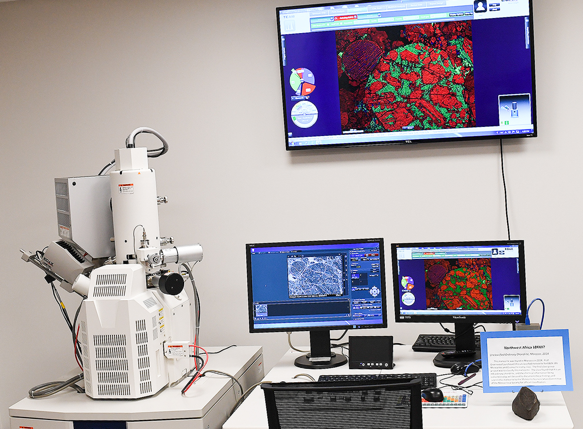
This February, the University replaced a 10-year-old scanning electron microscope (SEM) with a brand new field-emission SEM (FESEM), which provides higher resolution images at a faster speed. Its acquisition was enabled by a $202,300 grant from the National Science Foundation (NSF), awarded to Wesleyan in August 2017.
Assistant Professor of Earth and Environmental Science Jim Greenwood and Assistant Professor of Chemistry Michelle Personick led the application process, with the support of other faculty members. It was through the NSF’s Major Research Instrumentation Programs that the University received the grant.
“A field-emission cathode in the electron gun of a scanning electron microscope provides narrower probing beams at low as well as high electron energy, resulting in both improved spatial resolution and minimized sample charging and damage,” reads PhotoMetric Inc’s description of the machine. “Compared with convention scanning electron microscopy, field emission SEM produces clearer, less electrostatically distorted images with spatial resolution down to 1 1/2 nanometers – three to six times better.”
This upgrade in quality will allow for more accurate analyses of sample composition. The new microscope is already being used in two courses, and there are plans to incorporate it into several more.
“The new microscope is very specialized, but also very versatile,” Director of Scientific Imaging Jeff Gilarde told the Wesleyan Newsletter. “Researchers from biology, neuroscience, geology, physics and other departments may find the microscope of value to their work.”
The new machine will create many new opportunities for research on campus. The limitations of the SEM meant that Personick’s research group had to conduct most of its imaging at Yale University, where only graduate students are permitted to operate the machine. Now, both undergraduate and graduate students have daily access to the FESEM, and their research no longer depends on biweekly trips to New Haven.
“[It] is a versatile tool that enables researchers to simultaneously obtain a wealth of information about a sample, including topography, composition and fine structure,” Personick explained. “It means we can see very complex features on the surfaces of our nanoparticles, which are critical to how the nanoparticles behave chemically. With the old SEM, these same surfaces would appear smooth even on very large nanoparticles.”
The new instrument belongs to Wesleyan’s Advanced Instrumentation Center’s Scientific Imaging Laboratory and is located next to Shanklin Laboratory on the basement level.
Sasha Cohen can be reached at srcohen@wesleyan.edu.