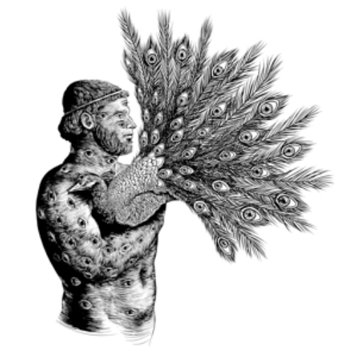
Since the early 1900s, fruit flies have been used as a model organism to study the relationship between an organism’s DNA and its appearance—that is, the relationship between genotype and phenotype. Early experiments quickly identified fruit flies’ potential to elucidate key genes in development. Altering these genes led to morphologically-dramatic mutants, such as fruit flies with legs growing from their heads or with an extra set of wings (both resulting from Hox gene mutations, if you’re interested).
The Johnson Lab at Wesleyan, spearheaded by Assistant Professor Biology Ruth Ineke Johnson, uses the same fruit fly organ used by Thomas Morgan in his research project in the 1910s that established fruit flies as the model organism for modern biology: the eye. While most fruit fly eyes are red, Morgan identified a few mutants with white eyes. By crossbreeding different fruit flies with each other, Morgan was able to see what factors led to inheritance of eye color, eventually leading to his postulation of the existence of chromosomes and linked traits, a fundamental theory in the field of molecular biology.
With modern advances in knowledge and technology, the Johnson Lab now uses the fruit fly eye in more complex ways and with more powerful tools. The Lab is interested in the way cells know where they should go in the body and how they know when they should die. This is especially important in both development and cancer. The developing embryo requires complex signaling pathways in order to develop properly. Interfering with the signaling pathway in any way can lead to dramatic phenotypic morphological effects, like the fruit flies with legs on their heads.
“If you would have asked me five years ago if I would end up studying cell death in the eye, I would have said no,” Johnson said. “I thought most of it had been worked out already. That is one of the joys of science. You follow the phenotypes and see where they take you.”
Cell death in development sounds like an oxymoron. It would seem that development is a process of creating new cells and sending them to the right places. Cell creation and motility is a large part of development, but programmed cell death plays an important role, as well. Many organs in the body develop with too many cells and knock off the excess cells in order to fine-tune the organ. But this process “costs energy,” as Johnson puts it. Understanding why, in the end, this process is more efficient and more effective than starting with the desired number of cells is one of the Johnson Lab’s ultimate goals.
The relationship between the study of development and the study of cancer is also an interesting one. On one hand, during development, cells divide more rapidly than they ever do in a mature organism. Thus, the pathways responsible for cell proliferation have also been linked to the cell proliferation observed in cancer. On the other hand, there is a fine-tuned mechanism in development that both prevents over-expression and general metastasis. That is, while the developing embryo’s cells are dividing more quickly and rapidly than in the developed organism, they do so with precise control over their rate of division and overall location within the embryo. Recently, the Johnson Lab has been looking at two different signaling pathways that are involved in determining whether a cell lives or dies in the fly eye.
In addition to building on Morgan’s precedent, the Johnson Lab uses the eye as a model for cell organization because of the fruit fly’s incredibly complex visual system. Henry Bushnell ’17 of the Johnson Lab paints a beautiful image of the eye, describing the eight hundred hexagons that fit together in a single eye as the “perfect lattice of a tiled floor.” Each hexagon is composed of 12 cells with different roles, organized in such a precise way as to allow it to fit together perfectly with the other 799 hexagons. This organization in the wild-type (genetically unchanged) eye is perfect to study what goes wrong when cells don’t know where to go or whether to live or die. It is obvious when a cell is out of place, as the entire lattice is disturbed.
After completing her undergraduate studies in South Africa, Johnson finished her PhD at the University of Cambridge. She came to the States for her post-doc at Washington University School of Medicine and Mount Sinai School of Medicine. While at Mount Sinai, Johnson discovered, characterized, and named the fruit fly protein “Cindr,” which she has continued to study at Wesleyan.
Cindr is an adaptor protein. Think of it like a matchmaker, the person in the room that drags their two friends together and helps them start a conversation. If the matchmaker protein weren’t present, molecule A and molecule B would never come together, but with Cindr’s help, they do. But it doesn’t just do this for A and B; It does it for a laundry list of proteins. After discovering Cindr, Johnson noticed an interesting protein that Cindr was shown to interact with: JNK.
JNK stands for Jun n-terminal kinase, which is actually a family of proteins that are all involved in the promotion of cell migration. This protein does not only exist in fruit flies; it also is observed in humans, though under a different name. JNK stood out for Johnson for its role in both development and cancer. In development, JNK is responsible for promoting the cell migration necessary for proper anatomical development. To highlight JNK’s role in development, scientists have eliminated certain JNKs to see what morphological changes occur. Normally, the adult fruit fly’s back tissue starts on its chest and extends like a coat of armor to connect in the back. But without JNK, the fruit fly’s chest begins extending but never fully closes, leading to flies with divots in their backs.
JNK is also active in the fully-developed organism. When a person gets a cut, JNK is responsible for migrating cells to fill in the chunk. Clearly, the over-expression of JNK could be problematic, hence its connection with cancer. The healthy cell, however, has tools to combat this problem. In fruit flies, if too much JNK is expressed, corresponding to excessive cell mobility, the cell automatically triggers its own death. Cancerous metastasis (malignant growths) has been associated with turning off this trigger for death. This is the story in fruit flies, but the story in humans is more complex.
The Johnson Lab works to study the interaction between Cindr and JNK by observing cell mobility and death in the developing eye. They use RNA-interference, a tool that takes advantage of the cell’s natural response to viral infection, to specifically control the amount of both Cindr and JNK present in the developing eye. The steps by which lab members observe the phenotypic effects of changes in Cindr and JNK concentration are the most fascinating.
“The pupa forms inside of a cocoon, which is like a sleeping bag,” Bushnell said. “You rip open a small hatch in the sleeping bag, take some small scissors, then snip along the sleeping bag seam, peel it back, and pull out the still alive pupa.”
The pupal brain is removed, which has two proto-eyes connected on either side.
“It looks rather like a dumbbell,” Johnson explained.
The eye-brain complex is then soaked in buffers containing compounds that specifically bind to molecules of interest to allow lab members to better visualize the organization of the cells in the eye. These compounds fluoresce at particular wavelengths of light, so with special microscopes, the lab can identify both protein location and general cell layout. The eye is observed under different microscopes that focus on the placement, size, number, and kind of cells in a single hexagon of the eye, to name a few.
Data published by the Johnson Lab in 2016 in “Developmental Biology” suggests a suppressive effect between Cindr and JNK in the fruit fly wing. That is to say, too much Cindr somehow downregulates the JNK pathway, leading to decreased cell mobility. This suggests a cancer-suppressing role of Cindr. The Lab is now working on observing the same effect in eye patterning.
“We worked out most of the details of the relationship [between JNK and Cindr] in the wing because it is the less complicated tissue,” Johnson said. “This didn’t give us the opportunity to study cell death. Now we’re going back and looking specifically in the eye to see how the relationship between Cindr and JNK is used to pattern the very organized organ.”
Beyond its practicality for cancer research, the work of the Johnson Lab is a step toward understanding the incredible rules that control complex organs. In short, they are searching for a rule book of development. Even though there may be different combinations of rules that pattern fruit fly organs as compared to human organs, the Johnson Lab seeks to understand the fundamentals and use them to generalize about the highly complex organisms that we see around us every day.


Leave a Reply